Female Internal Reproductive Organs Anatomy - Section 2 The Female Genital Organs Ppt Download - The female reproductive anatomy includes both external and internal structures.
byJordan Hurst•
0
Female Internal Reproductive Organs Anatomy - Section 2 The Female Genital Organs Ppt Download - The female reproductive anatomy includes both external and internal structures.. The female anatomy consists of female parts, which are external and internal. Female reproductive organs undergo substantial structural and functional changes every month. Looking at the anatomy of the internal structures of the female reproductive system, i've split this into two parts. The ovaries are the primary organs of the female reproductive system. Anatomy of female reproductive organs 1.
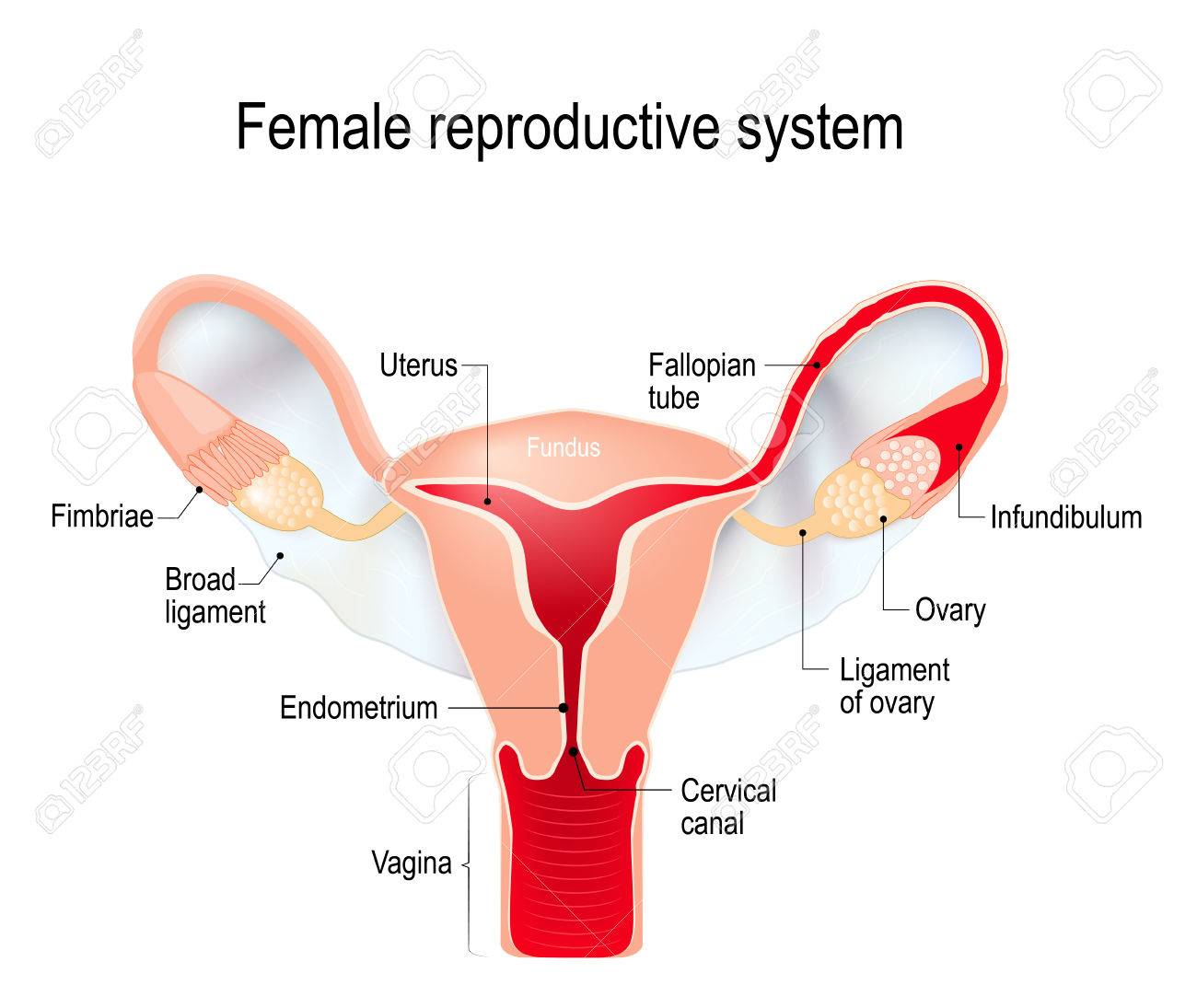
• serves as canal for menstrual fluid • forms the inferior part of birth canal. The ovaries are homologous to the testes of males bt lie within the pelvis. The passageway between the outside of the female body and the uterus. It produces the female egg cells necessary for reproduction, called the ova or oocytes. 2.5 cm • communicates superiorly with cervical canal and inferiorly with vestibule of vagina.
Labeled Female Reproductive System Diagram Jpg The Oncofertility Consortium from oncofertility.msu.edu Female internal organs anatomy human anatomy parts infographic. Fallopian tubes and ovaries form the adnexa of the uterus. An female's internal reproductive organs are the vagina, uterus, fallopian tubes, cervix, and ovary. The female internal reproductive organs are composed of the vagina, cervix, uterus, fallopian tubes, and ovaries. It is made up of the vulva, the vagina, the cervix, the uterus, the fallopian tubes and the ovaries. It produces the female egg cells necessary for reproduction, called the ova or oocytes. The ovaries are smaller than the testes. Female organs picture of female reproductive system diagram 1024×1204.
It also is known as the birth canal.
That being said, any part of your body can be sexual. The uterus (womb) is the site of implantation of a fertilized ovum, growth and development of the fetus during. The ovaries are the primary organs of the female reproductive system. Located at the top of the vagina, this is the lower most part of the uterus. It produces the female egg cells necessary for reproduction, called the ova or oocytes. • serves as canal for menstrual fluid • forms the inferior part of birth canal. The female internal reproductive organs are the vagina, uterus, fallopian tubes, and ovaries. Anatomy of female reproductive organs 1. It is made up of the vulva, the vagina, the cervix, the uterus, the fallopian tubes and the ovaries. The ovaries are within the pelvis, just lateral to the uterus. These organs are supported in the pelvis by ligaments. Female anatomy includes the external genitals, or the vulva, and the internal reproductive organs, which include the ovaries and the uterus. The female internal reproductive organs are composed of the vagina, cervix, uterus, fallopian tubes, and ovaries.
You will learn more about the overall anatomy of the female reproductive system at the end of this section. One major difference between males and females is their. Top view of red paper cut female reproductive internal organs with blood drops and menstrual cup on pink background medical exam. The uterus or womb accommodates the embryo which develops into the foetus. It serves important functions during pregnancy and childbirth.
Female Reproductive System Internal Sex Organs Uterus With Royalty Free Cliparts Vectors And Stock Illustration Image 73031134 from previews.123rf.com It is made of muscle and can stretch and grow. Anatomy of female reproductive organs 1. The female internal reproductive organs are the vagina, uterus, fallopian tubes, and ovaries. Located at the top of the vagina, this is the lower most part of the uterus. The ovaries are homologous to the testes of males bt lie within the pelvis. The main external structures of the female reproductive system include: The uterus is one of the predominant organs of the female reproductive system. The female reproductive tract is all located within the pelvis.
Female organs picture of female reproductive system diagram 1024×1204.
Female reproductive organs undergo substantial structural and functional changes every month. The interior membrane that lines the uterus is called. The uterus (womb) is the site of implantation of a fertilized ovum, growth and development of the fetus during. The uterus or womb accommodates the embryo which develops into the foetus. The female internal reproductive organs are composed of the vagina, cervix, uterus, fallopian tubes, and ovaries. The function of the external female reproductive structures (the genital) is twofold: The ovaries are within the pelvis, just lateral to the uterus. Learn about the placement of the skeletal and muscular structures. Diagram internal female anatomy : Reproductive organs include things like the uterus and testicles. Let's cover the major organs of the pelv. The female internal reproductive organs are the vagina, uterus, fallopian tubes, and ovaries. The female reproductive anatomy includes both external and internal structures.
This complex mechanism is also known as the. • serves as canal for menstrual fluid • forms the inferior part of birth canal. An female's internal reproductive organs are the vagina, uterus, fallopian tubes, cervix, and ovary. These organs are supported in the pelvis by ligaments. You will learn more about the overall anatomy of the female reproductive system at the end of this section.
The Female Reproductive System Boundless Anatomy And Physiology from s3-us-west-2.amazonaws.com The female reproductive tract is all located within the pelvis. Browse our female anatomy diagram organ images, graphics, and designs from +79.322 free vectors graphics. The ovaries are within the pelvis, just lateral to the uterus. Female reproductive organs undergo substantial structural and functional changes every month. The passageway between the outside of the female body and the uterus. Female reproductive organs undergo substantial structural and functional changes every month. Female organs picture of female reproductive system diagram 1024×1204. 2.5 cm • communicates superiorly with cervical canal and inferiorly with vestibule of vagina.
The ovarian cycle is a set of predictable changes in a female's oocytes and ovarian follicles.
The ovaries are homologous to the testes of males bt lie within the pelvis. Its anatomical structure can be broken down further into the mons pubis. The female internal reproductive organs are the vagina, uterus, fallopian tubes, and ovaries. It is made of muscle and can stretch and grow. The interior membrane that lines the uterus is called. That being said, any part of your body can be sexual. The female reproductive anatomy includes both external and internal structures. In virgins, the hymen usually encircles the opening like a tight ring, but it may completely cover the opening. This complex mechanism is also known as the. The passageway between the outside of the female body and the uterus. Browse our female anatomy diagram organ images, graphics, and designs from +79.322 free vectors graphics. Top view of red paper cut female reproductive internal organs with blood drops and menstrual cup on pink background medical exam. It produces the female egg cells necessary for reproduction, called the ova or oocytes.
The uterus is one of the predominant organs of the female reproductive system female internal. This complex mechanism is also known as the.